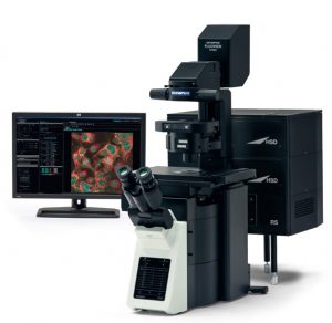
Laser scanning confocal microscope
- Name: Laser Scanning Microscope
Laser scanning microscopy is used in biological and materials science research to obtain high-resolution, high-contrast imagery of a sample. Laser microscopes can scan samples point by point, resulting in optical sectioning that can be used to construct precise 3D imagery. These microscopes obtain high-contrast three-dimensional images of microscopic structures by detecting the small amounts of fluorescent light emitted by a sample as it is scanned by a laser beam.
Instrument name: Laser scanning confocal microscope
Model: FV3000
Manufacturer: Olympus
Microscope Frame: Inverted microscope
Objectives: 4X, 10X, 20X, 40X, 60X (oil immersion), 100X (oil immersion)
Lasers: 405 nm, 488 nm, 561 nm, 640 nm
Dyes: Preloaded
Sample thickness: 200µm (max)
Spatial resolution: Along xy – 250 nm, Along z – 500 nm
Neutral density filter: ND-10
Scanner:
- Galvanometer scanner (Normal imaging)
- Resonant scanner (High speed imaging)
Spectral detector:
- Spectral detector (SD): Multi-Alkali photomultiplier, 2 channels
- High sensitivity spectral detector (HSD): Cooled GaAsP photomultiplier, 2 channels
Holders: Microplate holder, Slide holder, Dish holder Image techniques: Confocal imaging, Time lapse, Z-stack, 3D video making, Maximum intensity projection, FRAP
Technical Documents
Status:
Operational
Sample requirement:
Solid, liquid, thin film
For 100x and 10x magnification sample need to be placed on cover slip
Sample size must be 1 cm x 1 cm
Location: MNCF, Bio wing
Training Videos: Confocal training video new.mp4
Lieca flourescence microscope
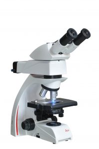
- Name: Lieca flourescence microscope
Fluorescence microscopy is a very powerful analytical tool that combines the magnifying properties of light microscopy with visualization of fluorescence. Fluorescence is a phenomenon that involves absorbance and emission of a small range of light wavelengths by a fluorescent molecule known as a fluorophore.
The Leica DM500 microscopes provide superior image quality along with durability and a practical design that truly meets the needs. The brightfield achromatic model offers 4x, 10x, 40x, and 100x plan objectives.
- Camera imaging system-Leica DFC450 C camera with C Mount 0.55X.
- Fluorescence illumination system- Fluorescent Filters sets: red, green and blue(A, I3,
- N2.1)
- 100W illumination is ideal for highly light absorbent specimens and DIC.
- 6-fold nosepiece
- Ergo stage with ceramic stage plate (left/right adaptation possible.)
- Transmitted light bright field (BF)
- Condenser achr.-apl. (P) with switchable condenser top and aperture diaphragm
- Planachromats N PLAN 5x, 10x, 20x, 40x, 100x, pair of eyepieces 10x/22M
Technical Documents
Status:
Operational
Sample requirement:
Solid, liquid, thin film
For 100x and 10x magnification sample need to be placed on cover slip
Location: MNCF, Bio wing
Training Videos:
BIOWING – Cancer biology lab
At MNCF – BIOWING, we are dedicated to pioneering advancements in cancer biology research. Our state-of-the-art facility is equipped with cutting-edge technologies and staffed by a team of passionate scientists committed to combating cancer through innovative research methodologies.
Our Services:
- Cell Culture: We specialize in the cultivation and maintenance of various cell lines, facilitating research into cancer cell behaviour and response to treatment.
- Cell Storage: Our lab provides secure and controlled storage solutions for precious cell samples, ensuring their integrity and viability for future studies.
- Preliminary Drug Testing: We conduct rigorous preliminary drug testing to evaluate the efficacy and safety of potential cancer therapeutics, contributing to the development of novel treatment strategies.
- Magnetic Hyperthermia: Utilizing innovative techniques, including magnetic hyperthermia, we explore non-invasive approaches to cancer treatment, aimed at targeted destruction of tumor cells while minimizing harm to healthy tissue.
- Autoclaving: With our advanced autoclaving facilities, we maintain strict sterilization protocols to guarantee the purity and safety of experimental materials and equipment.
- Other instruments are Centrifuge, water bath, C02 incubator, inverted microscope, hot air oven.
Our Mission:
At MNCF – BIOWING, our mission is to unravel the complexities of cancer biology and translate scientific discoveries into tangible advancements in cancer treatment and patient care. Through collaboration, innovation, and relentless dedication, we strive to make meaningful contributions to the global fight against cancer.
For more details, contact us.